Cranial Nerves & The Special Senses
Cranial nerves are the pathways to perception of
•Olfaction
•Taste
•Visual system
•Hearing and balance
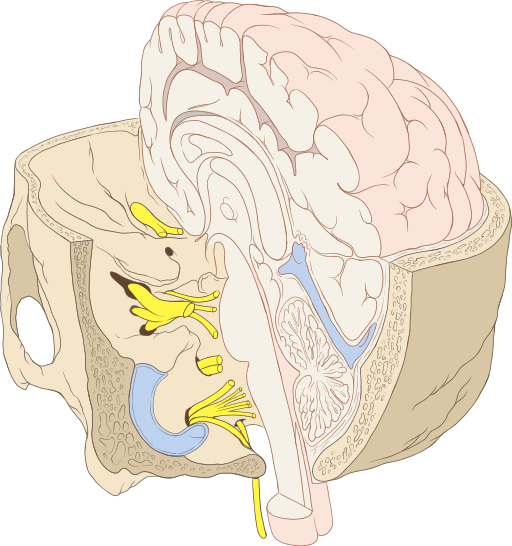
Cranial nerves reside in throughout the brain: cerebrum, and brain stem (midbrain, pons, and medulla oblongota). PTs will screen and assess gross cranial nerve function through observation of involuntary movement and simple motor commands.
Brainstem: connects spinal cord to brain; integration of reflexes necessary for survival

By Patrick J. Lynch, medical illustrator (Patrick J. Lynch, medical illustrator) [CC-BY-2.5 (http://creativecommons.org/licenses/by/2.5)], via Wikimedia Commons
Cranial nerves (CN): part of peripheral nervous system which arises directly from brain. Two pairs of CN arise from cerebrum; ten pairs arise from brainstem. Indicated by
Roman numerals I-XII from anterior to posterior. CNs may have one or more of three functions
- Sensory (special or general)
- Somatic motor (control of skeletal muscles)
- Parasympathetic (regulation of glands, smooth muscles, cardiac muscle)
Subdivisions and Associated Cranial Nerves
|
Brain Subdivision |
Associated Cranial Nerve |
Major Function |
|
cerebrum |
I: Olfactory |
Smell and taste |
|
thalamus |
II: Optic |
Vision |
|
midbrain |
III: Oculomotor IV: Trochlear |
Eye movements (adduction, elevation, depression) and motor for eyelid |
|
pons |
V: Trigeminal VI: Abducens VII: Facial VIII: Vestibulocochlear(part) |
Face sensation and motor Lateral eye movement Face sensory and motor, taste
Hearing and balance |
|
medulla oblongata |
VIII: Vestibulocochlear (part) IX: Glossopharyngeal X: Vagus
XI: Spinal Accessory XII: Hypoglossal |
Hearing and balance
Taste posterior third of tongue and sensory/motor in throat Autonomic functions for viscera, (BP, HR, RR), speech, breathing Motor to SCM and trapezius Motor function for tongue |
Cranial Nerve Tests and Measures
The Wayne State Tutorial is highly detailed and includes a section on "interpretation" which is outside of the PTA scope of practice. We have narrowed the focused of the tutorial by linking the procedures for testing in the table below.
Any patient who reports and/or demonstrates new signs and symptoms of cranial nerve dysfunction may be experiencing a medical emergency.
A PTA should be able to select and perform tests for cranial nerve function. Specific cranial nerve testing procedures are linked below:
|
Cranial Nerve |
Procedures |
Video Demo of Normal Function |
|
I - Olfactory |
||
|
II - Optic |
||
|
III, IV, VI - Eye control |
||
|
V - Trigeminal |
||
|
VII- Facial |
||
|
VIII - Vestibulocochlear |
||
|
IX, X - Gag Reflexes |
||
|
XI - Spinal Accessory |
||
|
XII - Hypoglossal |
For your reference, you can link to the full Wayne State Cranial Nerve Tutorial
Abnormal Cranial Nerve Tests and Measures
Abnormal Cranial Nerve II
Recall that visual fields are dependent on individual eye function as well as convergence of both eyes on an object/image. Damage to central visual processing systems can result in visual disturbances or field cuts.
|
Full Visual Field - Binocular Vision |
Left Visual Field Cut |
|
|
|
http://en.wikipedia.org/wiki/Visual_field
The video provides an example of a patient with a R visual field cut.. No sound
This patient demonstrates unilateral sensory dysfunction in the Trigeminal nerve. Recall that the the Trigeminal nerve is the primary nerve anesthetized during dental procedures.
Abnormal CN VII: Note the asymmetry in facial muscles
The tongue will deviate toward the affected (weaker) side when the patient is prompted to stick out their tongue
Cranial nerve coordination examination
Sample of Normal VOR
The VOR is also referred to as the "Doll's Eye's" reflex. The examiner (sound only) is assessing both eye motor function and coordination of eye and head motions which includes CN VIII
Safety Considerations
Examples of possible safety considerations for patients with CN deficits include:
- placing yourself and transfer surfaces within the patients's intact visual field; securing call bell within visual and sensory fields
- orienting the patient to engage in compensatory strategies to accommodate for losses in vision, sensation, etc.
- checking swallowing precautions prior to offering any patient with a neurological condition food/drink
- obtaining a second person for assistance as needed when there is a balance disorder
- applying/utilizing adaptive equipment during therapy based on deficits (eye patch, written/visual instructions (e.g., with hearing loss), soft-restraint to prevent injury to self, positioning/splinting to challenge or rest affected structures.


