

Applying Anatomical Planes and Direction of Motion
Describe normal movement and abnormal movement conditions, such as restricted movement or contracture, is based in a uniform use of anatomical position, planes, and axes of motion. As mentioned in our discussion of body mechanics, there are three anatomical reference planes: sagittal, frontal, and transverse (horizontal). There are three reference axes in each plane that are used to describe motion: frontal, sagittal, and vertical:
- Flexion / extension: Movement occuring in a sagittal plane across a horizontal axis
- Abduction / adduction: Movement occuring in a frontal plane across a sagittal axis
- Rotation (medial/lateral or external/internal): Movement occuring in a transverse (horizontal) plane across a vertical axis.
![]()
![]()
As you review the images and descriptions of the common contractures below, note the contracture is named based on the direction of the motion restriction.
Common Contractures
|
Area |
Clinical Considerations |
Image |
|
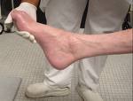
ankle plantar flexion |
prevented or minimized with braces, splint, weight bearing (e.g., standing) and habit modification (footwear)
|
|
|
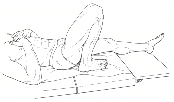
hip and knee flexion |
generally initiated with prolonged positioning with pillows under knees if patients are using a gatched bed (mattress bows at pelvis with HOB and feet elevated), risk for hip flexion contracture increases certain neurological conditions will cause the hip and knee to pull into flexion when at rest
|
|
|
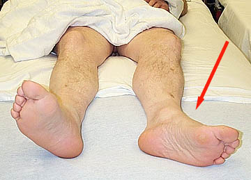
Hip external rotation |
External rotation is the "open" position of the hip joint. When there is significant extremity weakness, external rotation tendencies are often difficult to avoid
|
https://instruct.uwo.ca/kinesiology/222/Lab6/Lab6_Images/Fig4a_hip_fracture_patient_leg_external_rotation.jpg |
|
Cervical flexion |
Patients are at risk cervical flexion contractures when positioned with too many pillows or with HOB > 30 degrees for extended periods. Some patients with swallowing restrictions or dysfunction, cardiac condition, lung condition can not safely lie flat. These patients have higher cervical flexion contracture risk
|
|
|
Wrist flexion |
increased spasticity of muscles from neurological disease or injury or general disuse generally accompanies finger and thumb flexion contractures
|
|
The Life of a Pressure Sore
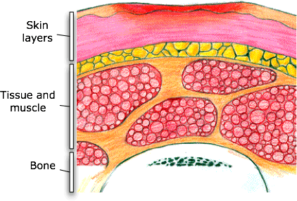
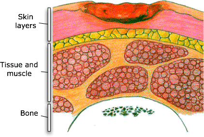
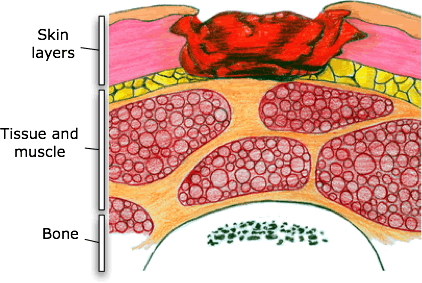
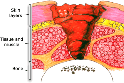
Pressure sores are noted and documented based on STAGES of tissue responses to pressure and shearing.
The two hour guideline for checking in with patients/residents and assisting with a change in position for pressure sore prevention is based on the amount of time it takes tissue to progress from Stage 1 to Stage 2 pressure sore.
|
Stage |
Description |
Onset |
Signs |
Resolution |
Image |
|
1 |
Non-blanching Hyperemia (redness that does not turn white when you push on it)
|
within 30 min
|
skin redness; patients with dark skin may have discoloration instead of redness
|
1 hour after removal of
|
|
|
2 |
Ischemia (decreased oxygen delivered) |
2-6 hours |
skin blanching (whiteness) |
36+ hours and after removal of pressure |
|
|
3 |
Necrosis (tissue death) |
6 hours |
skin blueness, hard lump |
varies (days/weeks) |
|
|
4 |
Ulceration |
Within 2 weeks of necrosis |
ulceration, infection, bony prominences involved
|
months; frequently requires surgical repair or amputation. May be fatal
|
|
Role of the PTA in Pressure Sore Treatment
A physical therapist evaluates and checks the skin conditions as part of routine care for clients under their care. PTs/PTAs check the epidermis, the dermis or the underlying area of tissue damage. This may include the muscles, tendons or bones. Wound care and dressing changes are within the scope of practice for PTA, however, treatment is complex. A PTA involved in direct wound care is likely to have significant clinical experience and additional continuing education in wound care
Wounds will not completely heal unless the underlying cause of the pressure sore is addressed. Reduction of pressure and prevention of future breakdowns are a PT/PTAs top priority. A procedure to assess the amount of weight bearing across the pelvis is called pressure mapping. Pressure mapping may be recommended to assess specific pressure loads when a patient is sitting in a wheelchair so an optimal wheelchair cushion and seat back can be identified. A wheelchair seating vendor, occupational therapist (OT), nursing and physician are often part of the health care team providing assessment and treatment for pressure sores.
