Cardiac Anatomy and Physiology
Back in PTA 100....
In Unit 3 of PTA 100, we covered the role of physical therapy with patients with cardiovascular conditions. We encourage you to go back to your notes from Unit 3. Specific examples of stress testing is included in this section. You should start to recognize that the interventions for conditioning and cardiac conditions are similar in that the primary impairment is typically impaired aerobic capacity and endurance.

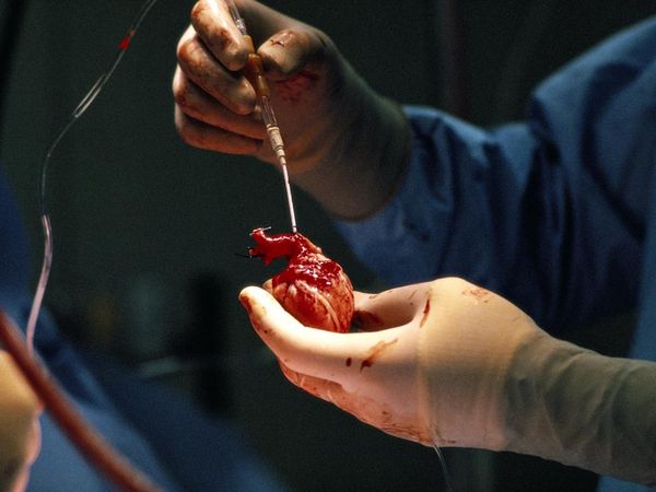
Donor Heart
The following information is all review from anatomy. Utilize it as personally needed.
Review anatomy of heart chambers, coronary arteries and heart conduction system in anatomy text. Tutorials to refresh your memory are included below:
You are not expected to memorize the values contained in the above link
Specific anatomical references in the heart are below. You are not expected to recall anatomy at this level. These resources are included for your reference only if you would like more specific landmarks for cardiac anatomical terms
External Anatomy of the Heart (ASPSU, 2019)
apex - The tip or pointed end of the heart; in the anatomical position, the confluence of the inferior and left borders; it is a projection inferiorly, anteriorly and to the left of the left ventricle.
base - The broad superior surface of the heart, from which the great vessels emerge.
auricle - (1) The free portion of the myocardial wall of each atrium which has a superficial similarity in shape to an ear, it contracts along with the rest of the atrial wall to move blood from the atria to the ventricles; it is regulated by the Autonomic Nervous System; (2) The outer projecting portion of the ear, also called the pinna.
coronary sulcus - The slight groove seen on the external surface of the heart which indicates the borders between the atria and the ventricles; coronary vessels follow portions of the groove.
interventricular sulcus - The slight groove seen on the external surface of the heart which indicates the borders between the right and the left ventricles; coronary vessels follow portions of the groove.
