

Neurological System Functions
Fun
In order to understand what can go wrong, we need to know what normal function looks like. It is easier to understand signs and symptoms of disease when you can reference the involved structures and processes. PT interventions, tests, and measures become more meaningful when you have a general understanding of deficits and potential for rehabilitation.
Functional Overview
1. Sensory: Monitor internal and external stimuli
2. Integration: Brain and spinal cord process sensory input and initiate responses
3. Control: Muscles and glands
4. Homeostasis: Regulate and coordinate physiology
5. Mental activity: Consciousness, thinking, memory, emotion

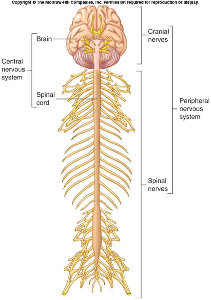
Organization of the Nervous System
Concepts and Terminology to Learn
- Peripheral Nervous System
- Central Nervous System
- Autonomic Nervous System
- Motor Neurons
- Sensory Neurons
Components on the Nervous System
–Brain, spinal cord, nerves, sensory receptors
Subdivisions
–Central nervous system (CNS): brain and spinal cord
–Peripheral nervous system (PNS): sensory receptors and nerves
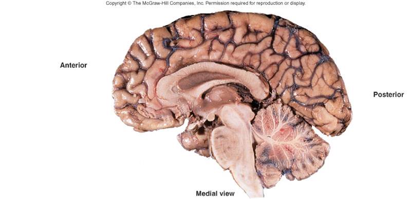
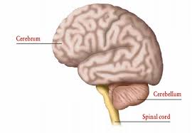
Brain
Brain
•Part of CNS contained in cranial cavity
•Control center for many of body's functions
Parts of the brain
•Cerebrum/cerebral cortex: conscious thought, control
•Brainstem: connects spinal cord to brain; integration of reflexes necessary for survival
•Cerebellum: involved in control of locomotion, balance, posture
•Diencephalon: thalamus, subthalamus, epithalamus, hypothalamus
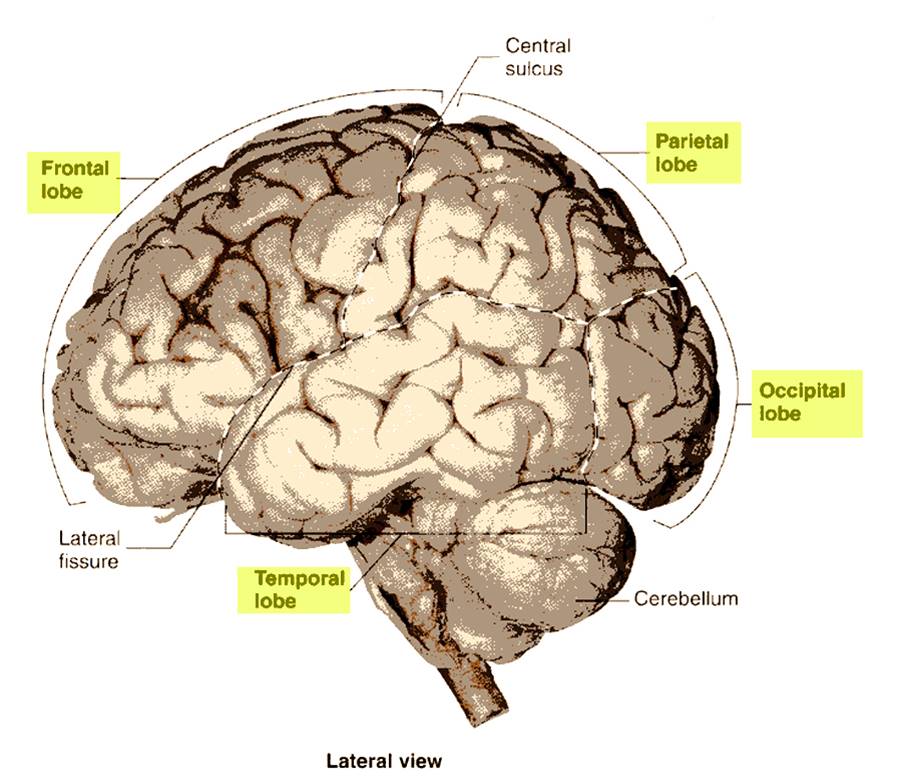
Cerebrum
•Largest portion of brain
•Composed of right and left hemispheres each of which has the following lobes: frontal, parietal, occipital, temporal, limbic, insular
Cerebral Lobes
•Frontal: executor of function: voluntary motor function, motivation, aggression, sense of smell, mood
•Parietal: sensory integrator for pain, temperature, detection of taste, and touch; coordinates reading
•Temporal: Reception and evaluation for smell and hearing; memory, abstract thought, judgment; Insula is within temporal lobe.
•Occipital: reception and integration of visual input
•Central sulcus: between the pre central gyrus/primary motor cortex and post central gyrus/primary somatic sensory cortex
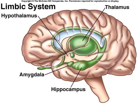
Limbic System
•Part of cerebrum and diencephalon
•Basic survival functions such as memory, reproduction, nutrition
•Emotions
•Various nuclei of the thalamus
•Part of the basal nuclei, hypothalamus, olfactory cortex, fornix
Diencephalon
- Connects the brain to the brainstem: image above
- Components: thalamus, subthalamus, epithalamus, hypothalamus
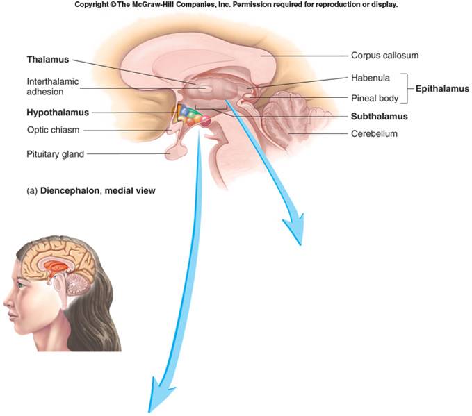
Thalamus
•Sensory information from spinal cord synapses here before projecting to cerebrum
•Relay information to motor, mood, emotion, and sensory integration areas in the cerebral cortex
Subthalamus
•Involved in controlling motor function
•Contains subthalamic nuclei, parts of red nuclei and substantia nigra.
•Several ascending and descending nerve tracts

Epithalamus
•Pineal gland
- –may influence sleepiness, helps regulate biological clock, may play a role in onset of puberty
- –Role in emotional and visceral responses to odors
Hypothalamus
•Most inferior portion of diencephalon
•olfactory reflexes and emotional responses to odors
•Controls endocrine system.
•Receives input from viscera, taste receptors, limbic system, nipples, external genitalia, prefrontal cortex
•Efferent fibers to brainstem, spinal cord (autonomic system), to posterior pituitary, and to cranial nerves controlling swallowing and shivering
•Important in regulation of mood, emotion, sexual pleasure, satiation, rage, and fear
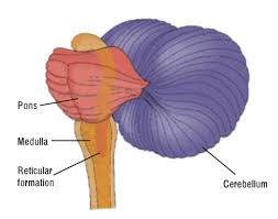
Brainstem
•Comprised of midbrain, pons, and medulla oblongata.
•Considered Peripheral Nervous System (PNS)
•These peripheral nerves originate from brain.
•Two pairs arise from cerebrum; ten pairs arise from brainstem
A pontine CVA is a stroke involving the brainstem.
•Continuous with spinal cord; has both ascending and descending nerve tracts
•Regulates: sleep, heart rate, blood vessel diameter, respiration, swallowing, vomiting, hiccupping, coughing, and sneezing

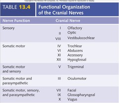
Crainial Nerves
- Considered Peripheral Nervous System (PNS)
- These peripheral nerves originate from brain.
- Two pairs arise from cerebrum; ten pairs arise from brainstem
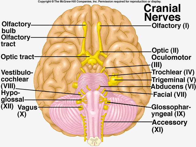
•Indicated by
- –Roman numerals I-XII from anterior to posterior
- –Names
•May have one or more of three functions
- –Sensory (special or general)
- –Somatic motor (control of skeletal muscles)
- –Parasympathetic (regulation of glands, smooth muscles, cardiac muscle)
Cranial Nerve Reflexes
- X (Vagus): reflexes having to do with heart rate, blood pressure, and respiration
- Reflexes involving both cranial nerves and brainstem:
- Turning the eyes toward sudden noise, touch on skin, flash of light
- Eyes tracking a moving object.
- Reflex using VIII, V, and VII to contract muscles associated with middle ear that protect ear ossicles
- Chewing reactions to textures of food, movement of tongue pushing food under tooth-row and out of harm's way
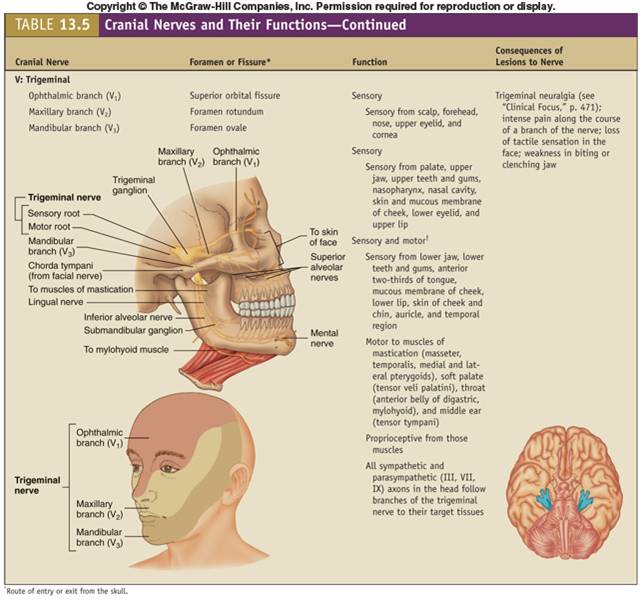
Trigeminal Nerve
Cerebellum
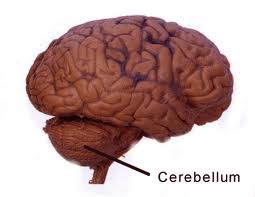
- •Key Point: COORDINATION; Ataxia = lack of coordination
- •Makes comparisons between the motor plan from the cortex and the position sense from the muscles/joints and facilitates movement precision/correction
- •Influences timing and force of voluntary muscular contraction
•Finger-to-nose test: what this is testing is coordination ruling out ataxia. If a pt had an impaired finger to nose test, the PTA would not interpret meaning, but give/chart this information as impaired or not impaired and share results with the supervising PT.
Cerebrum versus Brainstem
- •Brainstem and diencephalon maintain homeostasis of basic/primitive functions
- •Cerebrum and cerebellum coordinate, plan, and memorize higher level sensori-motor function
The two systems interact in automatic and conscious ways throughout the life cycle
Blood Supply to the Brain
![]()
![]()
•Anterior Cerebral Artery (ACA)
- Frontal, parietal, and basal ganglia
•Middle Cerebral Artery (MCA)
- Lateral surfaces of the frontal, parietal, temporal and occipital lobes
•Posterior Cerebral Artery (PCA)
- Midbrain, thalamus, occipital lobe, medial and inferior temporal lobe
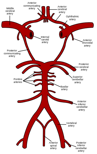
Image is of the various arteries that supply blood to the brain
Basilar Artery
–Includes anterior inferior cerebellar artery (AICA), superior cerebellar artery
–Supplies pons and cerebellum
–Primary blood supply to midbrain
–Complete occlusion can be fatal
Circle of Willis
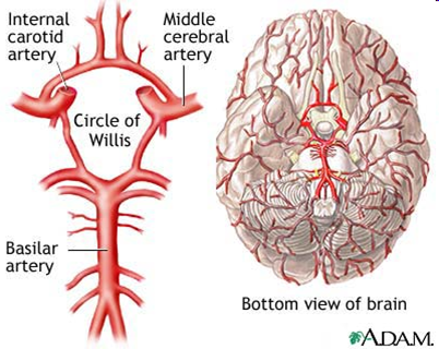
•Ring of 9 arteries
•Provides multiple sources of circulation/blood supply to the cerebrum
Vertebral Arteries
–Carry one-third of blood supply to the brain
–Originate from the subclavian artery
–Branches into three parts
- •Anterior Spinal
- •Posterior Spinal
- •Posterior Inferior Cerebellar Artery (PICA)
–All three branches supply blood to medulla
–PICA supplies inferior cerebellum
Internal Carotid Arteries
–Originate from the common carotid
–Becomes the posterior communicating arteries (PCA)
–Divides into anterior and middle cerebral arteries
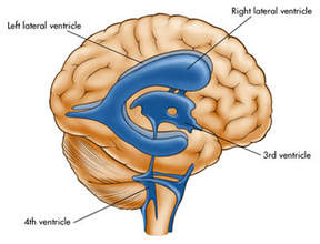
Ventricles
![]()
![]()
Ventricles are interconnected by aqueducts and wall openings
Blockage in the central canal or fourth ventricle can lead to hydrocephalus (enlarging ventricles) and may require an external shunt for treatment
Cerebrospinal Fluid (CSF)
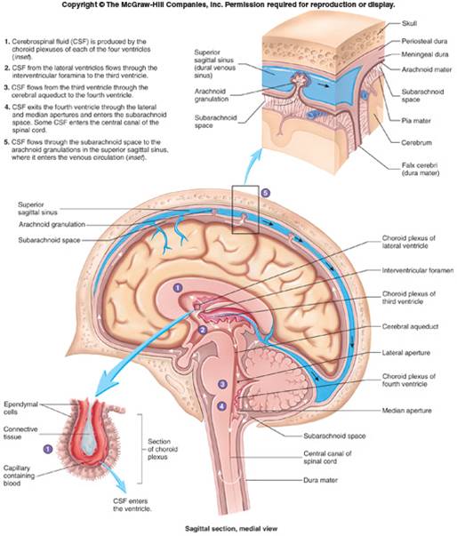
CSF
Similar to serum, but most protein removed
- •Bathes brain and spinal cord
- •Protective cushion around CNS
- •Choroid plexuses produce CSF which fills ventricles and other parts of brain and spinal cord
Composed of ependymal cells, their support tissue, and associated blood vessels
Blood-cerebrospinal fluid barrier
- •Endothelial cells of capillaries attached by tight junctions
- •Substances do not pass between cells
- •Substances must pass through cells
- •Makes the barrier very selective
