Electrical Stimulation for Tissue Repair
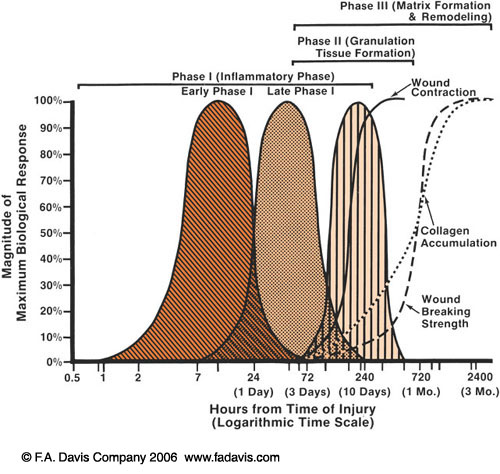
Let's recap the phases of wound healing quickly. Remember that there is a predictable sequence to tissue healing but the phases also overlap.

Phase 1: Inflammatory Phase
- Up to 3-7 days post insult
- Immediate vasodilation to protect the area followed by localized vasodilation to bring in the "clean up crew" macrophages via chemical signal or "chemotaxis"
Phase 2: Proliferation (granulation) Phase
- 3 days - 1 month post insult
- Multiple events occurring:new blood cells forming, extracellular matrix to fill space and form granulation tissue as scar tissue foundation, FIBROBLASTS are busy building new tissue, then wound contracts to pull edges back together
Phase 3: Remodeling Phase
- Can start within a few days and last up to 2 years following wound closure
- Significant increase in collagen, converting and reorganizing into strong, more elastic scar
How can electrical stimulation affect wound healing?
"Current of Injury" occurs when there is interruption of the normal skin barrier with negative outer layer (stratum cornue m) and positive inner dermis layers, creating a voltage gradient at the edge of the wound. This positive polarity flows out from the wound and returns via the sodium potassium pump.
- Electrical stimulation may mimic the normal body electric current to facilitate the wound healing process, though not determined
- Wound polarity is initially positive for the first 3-4 days then switches to negative, a sign of healing
"Galvanotaxis" - electrodes act to carry charges cells within the wound since cells are attracted to either a positive or negative pole based on their own opposite positive or negative charge
Indications: wounds from the following categories:
- Pressure (ducubitus) ulcers
- Venous insufficiency
- Arterial insufficiency
- Diabetes Mellitus
Contraindications
- Demand cardiac pacemakers
- Over the carotid sinus
- Site of skin lesions: basal cell carcinoma, squamous cell carcinoma, melanoma
- Osteomyelitis directly under the wound
- Presence of metal from antimicrobial medication or implants
- Lumbar region over pregnant woman
Precautions
- Wounds located at the cranium or upper cervical area
- Patient history of epilepsy, CVA, or reversible ischemic neurological deficiency

Application Techniques
- Direct Technique - Active electrode placed directly over wound with dispersive 15-20cm distal to wound
- high voltage monophasic pulsed current
- saline soaked gauze in wound bed for debridement
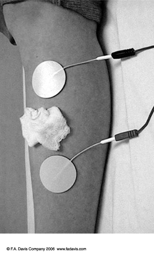
2. Periwound Technique - 2 electrodes are placed on intact skin adjacent to wound
- generall proximal and distal, but can be lateral as well.
- less risk of skin/wound disruption
*See Application Checklist and Patient Instructions in Behren's text
